Bones In Leg Diagram - Foot Anatomy Foot Ankle Lower Leg Orthopedic Assessment - With different grades of sprains depending on severity.. These are the femur, patella, tibia, fibula, tarsal bones, metatarsal bones, and phalanges (see figure 6.51). High resolution textures and displacement included. It is likely that abnormal biomechanical stresses are the basis for the disease. The bones together make up the hip. They allow you to move and provide support for your upper body.
Ankle & lower leg anatomy. These are the femur, patella, tibia, fibula, tarsal bones, metatarsal bones, and phalanges (see figure 6.51). Dog leg anatomy is complex, especially dog knees, which are found on the hind legs. The biceps is attached to the arm bones by.biceps femoris, semitendinosus, and semimembranosus. Posted on april 18, 2019april 18, 2019.
Medicine Anatomy Skeleton Bones Joints Of The Human Arm Bones And Leg Bones Wood Engraving 19th Century 19th Century Graphic Graphics Brachial Joint Leg Joint Leg Joints Arm Bones Arm Bone from c8.alamy.com The bones of the leg are the femur, tibia, fibula and patella. Diagramme schnell und einfach erstellen. Distal end of right humerus. These are the femur, patella, tibia, fibula, tarsal bones, metatarsal bones, and phalanges (see figure 6.51). The medial, larger bone of the lower leg. Leg bones diagram diagram schematic ideas lower leg muscle diagram blank sketch coloring page antique 1890s medical anatomy diagram leg bones skeleton posted on april 18, 2019april 18, 2019. Your legs are two of your most important body parts. The upper leg, in particular, is comprised of bones and muscles that are susceptible to injury, particularly when excess strain is placed upon them.
Image result for leg bones diagram human leg bone structure your leg bones are the longest and strongest bones in your body.
Degenerative disease, similar to arthritis. The hip itself is a ball and socket joint, much like the shoulder.the structures necessary to create this joint are the socket, the joint capsule, muscle, ligaments, and the neck. The technical term for a dog knee is the stifle joint. The proximal portion of the tibia is tibial plateau which acts as a cusp for the knee, the distal portion tapers into the medial malleoli and the concave surface which articulates with the talus at the ankle joint. Leg bones diagram diagram schematic ideas lower leg muscle diagram blank sketch coloring page antique 1890s medical anatomy diagram leg bones skeleton posted on april 18, 2019april 18, 2019. Your legs are two of your most important body parts. Its decrease finish helps create the knee joint. These are the femur, patella, tibia, fibula, tarsal bones, metatarsal bones, and phalanges (see figure 6.51). Ankle & lower leg anatomy. This area is commonly referred to as the calf. Dog leg anatomy is complex, especially dog knees, which are found on the hind legs. Hip and leg bone diagram : You'll learn about the muscle.
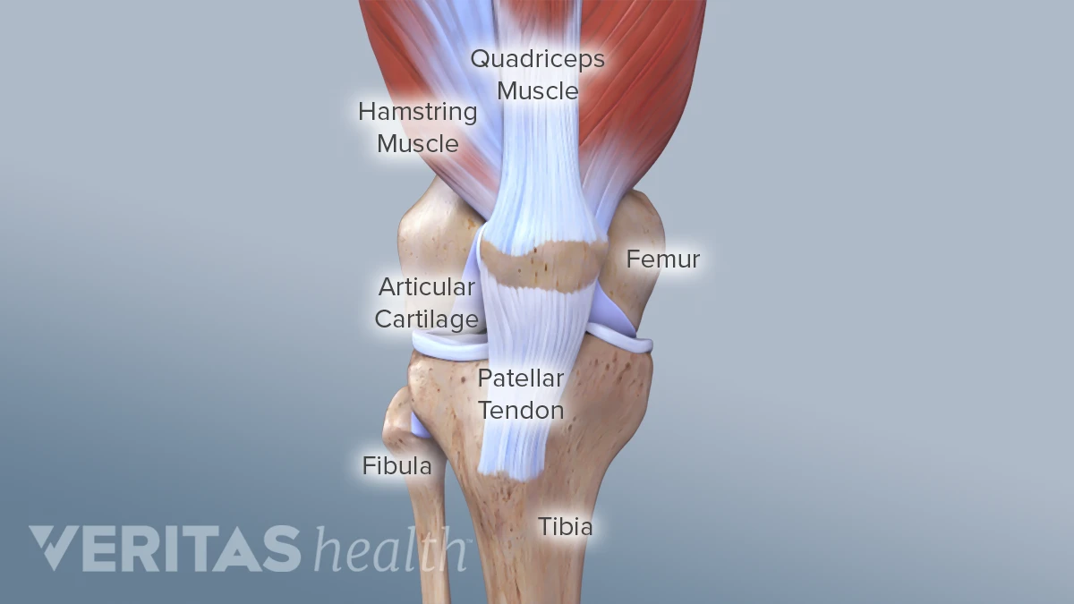
Pin on medical websites we like. The patella is the kneecap and articulates with the distal femur. The thigh bone, or femur, is the large upper leg bone that connects the lower leg bones (knee joint) to the pelvic bone (hip joint). Also called the shin bone, the tibia is the longer of the two bones in the. This area is commonly referred to as the calf.
Knee Anatomy from embed.widencdn.net They allow you to move and provide support for your upper body. Your legs are two of your most important body parts. The bones of the hip include the femur, the ilium, the ischium, and the pubis. The majority of muscles in the leg are considered long muscles, in that they stretch great distances. Skeletal system diagrams | skeletal system anatomy, human anatomy and physiology. Distal end of right humerus. The medial, larger bone of the lower leg. At the same time, the bones and joints of the leg and foot must be strong enough to support the body's weight while remaining.
The smaller lateral bone of the lower leg.
The upper leg, in particular, is comprised of bones and muscles that are susceptible to injury, particularly when excess strain is placed upon them. With different grades of sprains depending on severity. Bones in leg diagram color the leg on the left side. High resolution textures and displacement included. Pin on medical websites we like. The pubis, ischium, and ilium together constitute the pelvis while the thigh bone is the femur. The bones of the leg are the femur, tibia, fibula and patella.the foot bones shown in this diagram are the talus, navicular, cuneiform, cuboid, metatarsals and calcaneus. The bones of the leg and foot form part of the appendicular skeleton that supports the many muscles of the lower limbs. This allows weight to be distributed either anteriorly or posteriorly throughout the foot. Dog leg anatomy is complex, especially dog knees, which are found on the hind legs. The bones of the leg are the femur, tibia, fibula and patella. The knee joint is the largest joint in the body and is primarily a hinge joint, although some sliding and rotation occur. Its lower end helps create the knee joint.
Related posts of diagram of leg bones inside of arm muscle and bone. Hip and leg bone diagram : File human arm bones diagram svg wikipedia. I think this was the case this is a detailed diagram of a horse's hoof. The lower leg extends from the knee to the ankle.
Scanning X Ray Image Of Lower Leg Bone Download Scientific Diagram from www.researchgate.net Bones in leg diagram color the leg on the left side. Human leg bone diagram : Hip and leg bone diagram : The majority of muscles in the leg are considered long muscles, in that they stretch great distances. The tibia and the fibula, at the top of the ankle joint. Jan 28, 2016 · as you can see in the diagram above, the lower leg and ankle is a complex system of muscles, tendons, and joints. The technical term for a dog knee is the stifle joint. The bones of the leg are the femur, tibia, fibula and patella.the foot bones shown in this diagram are the talus, navicular, cuneiform, cuboid, metatarsals and calcaneus.
Diagram of blood and nerve supply to bone.
The bones of the hip include the femur, the ilium, the ischium, and the pubis. The knee joint is the largest joint in the body and is primarily a hinge joint, although some sliding and rotation occur. They allow you to move and provide support for your upper body. The major bones of the leg are the femur (thigh bone), tibia (shin bone), and adjacent fibula, and these are all long bones.the patella (kneecap) is the sesamoid bone in front of the knee.most of the leg skeleton has bony prominences and margins that can be palpated and some serve as anatomical landmarks that define the extent of the leg. The foot bones shown in this diagram are the talus, navicular, cuneiform, cuboid, metatarsals and calcaneus. The human leg consists of 8 bones, 4 per leg. Tibia and fibula the tibia and fibula are two long bones that run parallel to each other, forming the scaffold of the leg and providing attachment points for many muscles. Leg bone diagram / bones of the human leg 17. These landmarks are the anterior superior iliac spine. Performance horses tend to suffer from this degenerative disease. Human leg bone diagram : Distal end of right humerus. Your legs are two of your most important body parts.


0 Komentar